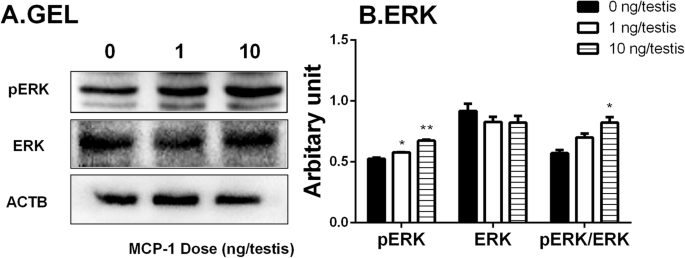
Morphology of Leydig cells in the testes after in vivo MCP-1 treatment.
Por um escritor misterioso
Last updated 16 outubro 2024

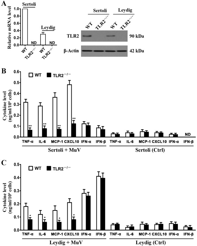
Mumps virus-induced innate immune responses in mouse Sertoli and Leydig cells

A brief exposure to cadmium impairs Leydig cell regeneration in the adult rat testis

Stem Leydig cells: Current research and future prospects of regenerative medicine of male reproductive health - ScienceDirect
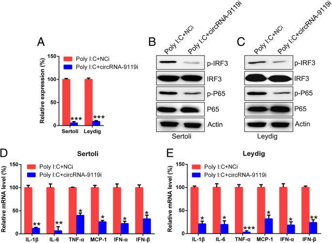
CircRNA-9119 suppresses poly I:C induced inflammation in Leydig and Sertoli cells via TLR3 and RIG-I signal pathways, Molecular Medicine
Frontiers Cytokines in Male Fertility and Reproductive Pathologies: Immunoregulation and Beyond

Morphology of Leydig cells in the testes after in vivo MCP-1 treatment.

Testicular macrophages are recruited during a narrow time window by fetal Sertoli cells to promote organ-specific developmental functions
IJMS, Free Full-Text
Monocyte Chemoattractant Protein-1 stimulates the differentiation of rat stem and progenitor Leydig cells during regeneration, BMC Developmental Biology
IJMS, Free Full-Text

PDF) Monocyte Chemoattractant Protein-1 stimulates the differentiation of rat stem and progenitor Leydig cells during regeneration
Cells, Free Full-Text
Recomendado para você
-
 Vivo NEX S mostra robustez em teste de durabilidade – Tecnoblog16 outubro 2024
 -
 Vivo realiza testes para diferentes aplicações do 5G no Rio de Janeiro16 outubro 2024
 -
 AO VIVO - Teste Seletivo SEMOSP16 outubro 2024
 -
 Governador de Nova York faz teste de coronavírus ao vivo pela TV – Política – CartaCapital16 outubro 2024
 -
/i.s3.glbimg.com/v1/AUTH_da025474c0c44edd99332dddb09cabe8/internal_photos/bs/2023/J/I/RRc9uPSAu4dWxgtdUqlg/tim.webp) TIM cria 'test-drive' para atrair clientes das rivais Claro e Vivo16 outubro 2024
TIM cria 'test-drive' para atrair clientes das rivais Claro e Vivo16 outubro 2024 -
 Internet movel ilimitada(teste gratis)c3 - Celulares e telefonia16 outubro 2024
 -
![Microsoft Azure DevOps - Foco em Testes Ágeis [Ao Vivo + On Demand] - Iterasys](https://cdn.eveclass.com/p/61cb81d816aa58f336ffe148/files/gallery/image/9e600cc0-fe01-11ec-a7d4-611f0e6ccdc5/thumbnail.jpg) Microsoft Azure DevOps - Foco em Testes Ágeis [Ao Vivo + On Demand] - Iterasys16 outubro 2024
Microsoft Azure DevOps - Foco em Testes Ágeis [Ao Vivo + On Demand] - Iterasys16 outubro 2024 -
 Vivo X90 Pro+ Review: Vivo sets the bar very high with its flagship smartphone - Reviews16 outubro 2024
 -
 Etilômetro testado ao vivo em repórter detecta álcool em ambiente; entenda, Ceará16 outubro 2024
 -
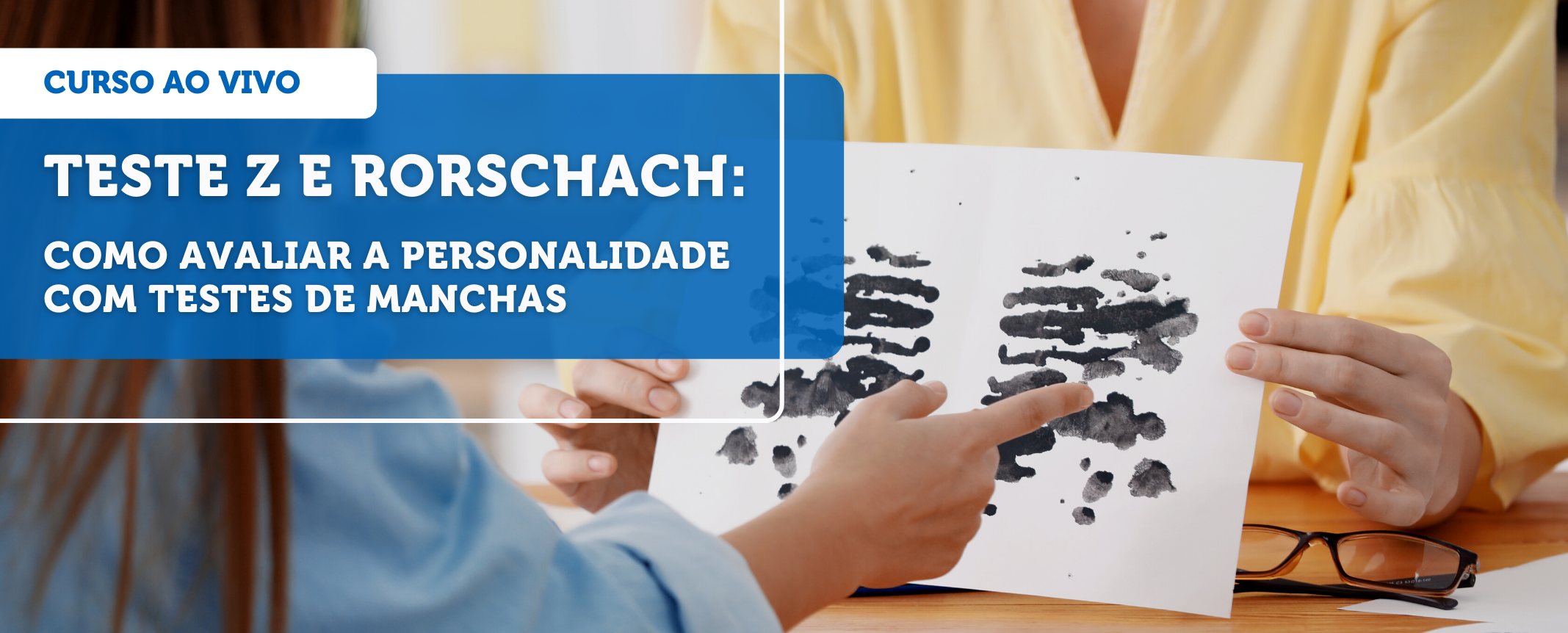 Rorschach e Teste Z: como avaliar a personalidade com testes de16 outubro 2024
você pode gostar
-
 Anime japonês kaneki ken topos y2k tóquio ghoul camiseta feminina punk harajuku dark cartoon tees streerwear S-2XL roupas femininas16 outubro 2024
 -
Instagram photo by IShowSpeed • Jul 21, 2021 at 5:57 PM16 outubro 2024
-
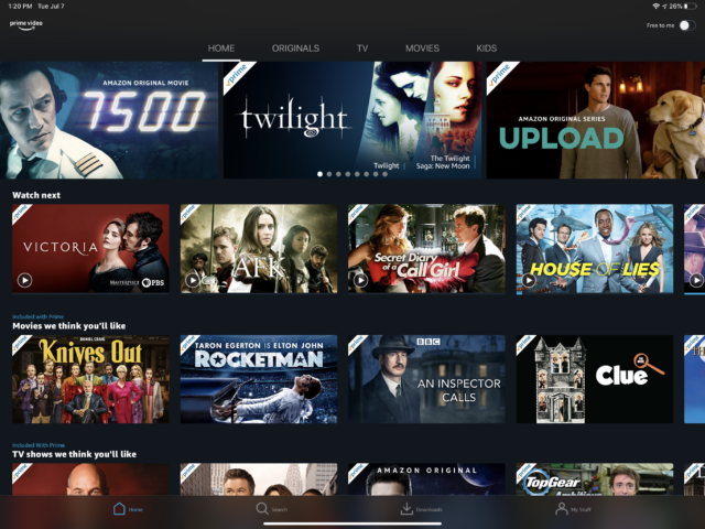 Prime Video will finally offer one of Netflix's most basic features16 outubro 2024
 -
 Jogo De Cordas Giannini Para Violão Aço Cobra 012 - Cordas para Violões - Magazine Luiza16 outubro 2024
 -
 WinKawaks » Roms » The King of Fighters '98: The Slugfest - The16 outubro 2024
 -
 Yoga in Pair. Women. Duo. Balance on One Leg Stock Photo - Image of concentration, background: 6495310816 outubro 2024
 -
 Classic Female v2 - Hair, Roblox Wiki16 outubro 2024
 -
 All Fist Skills in Project Mugetsu #roadto10k #roblox16 outubro 2024
 -
 Assistir 9-1-1 Online em Português - Pobre TV16 outubro 2024
 -
 Assassin's Creed Valhalla The Twilight Pack Xbox Key16 outubro 2024
