Typical magnetic resonance imaging scan showing the coracohumeral
Por um escritor misterioso
Last updated 22 janeiro 2025


Pain related to rotator cuff abnormalities: MRI findings without clinical significance - Bencardino - 2010 - Journal of Magnetic Resonance Imaging - Wiley Online Library
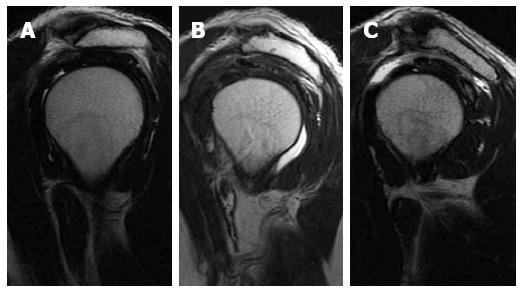
Rotator cuff disorders: How to write a surgically relevant magnetic resonance imaging report?

The Rotator Interval: A Review of Anatomy, Function, and Normal and Abnormal MRI Appearance

MRI of upper limb: normal anatomy

Magnetic Resonance Imaging (Part IV) - Clinical Emergency Radiology

BIR Publications

The Primer for Sports Medicine Professionals on Imaging: The Shoulder - Nadja A. Farshad-Amacker, Sapna Jain Palrecha, Mazda Farshad, 2013
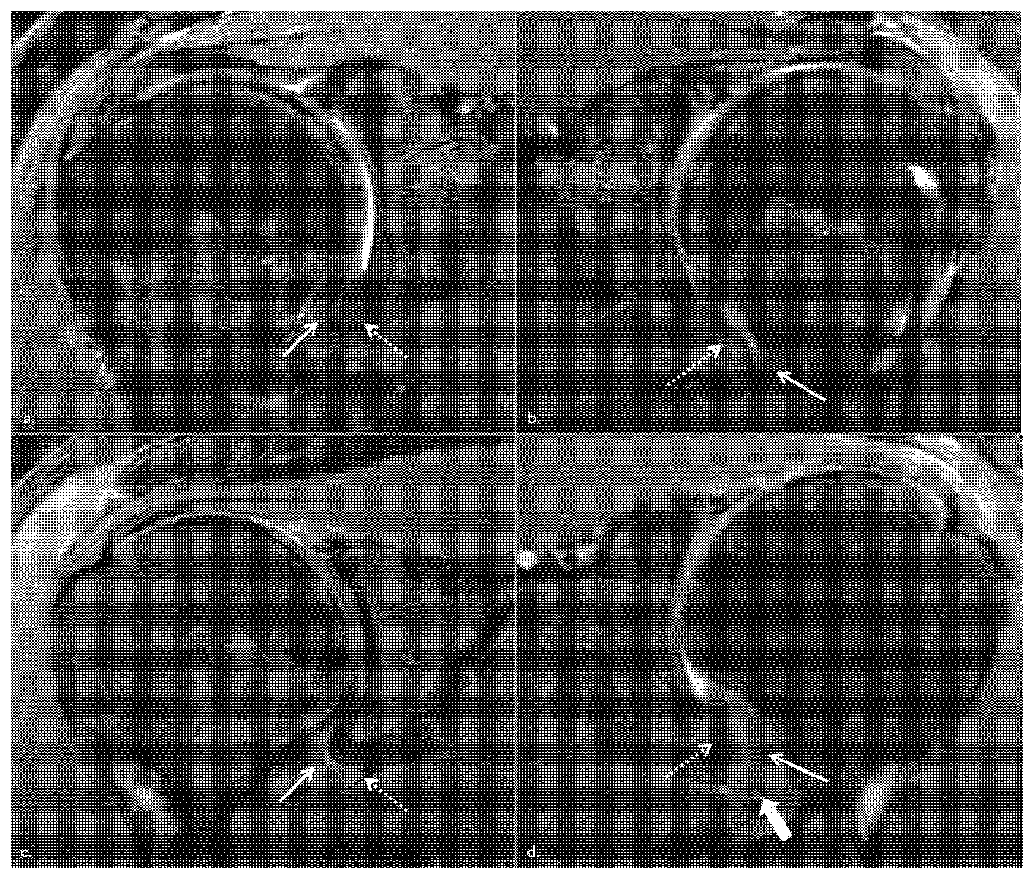
JCM, Free Full-Text

Magnetic resonance imaging of the shoulder
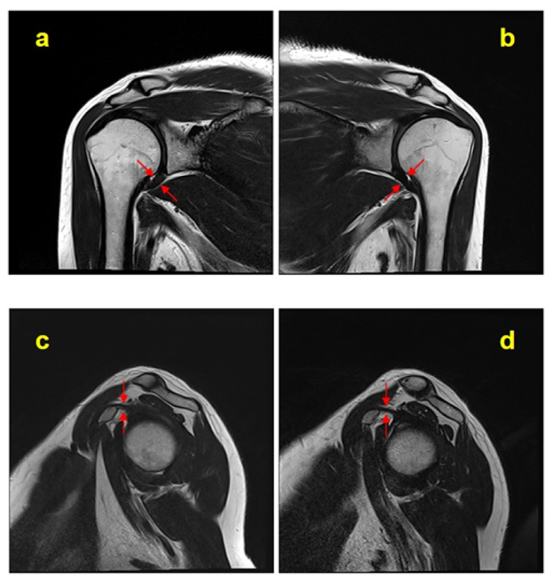
Contrast-enhanced Magnetic Resonance Imaging Revealing the Joint Capsule Pathology of a Refractory Frozen Shoulder

Presentation1, radiological imaging of adhesive capsulitis(frozen shoulder).

Glenohumeral Instability

A Systematic Approach to Magnetic Resonance Imaging Interpretation of Sports Medicine Injuries of the Shoulder - Timothy G. Sanders, Mark D. Miller, 2005
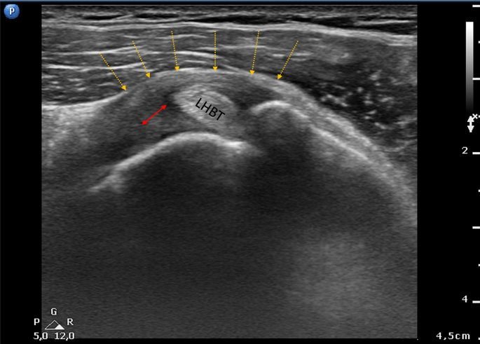
Ultrasound Features of Adhesive Capsulitis
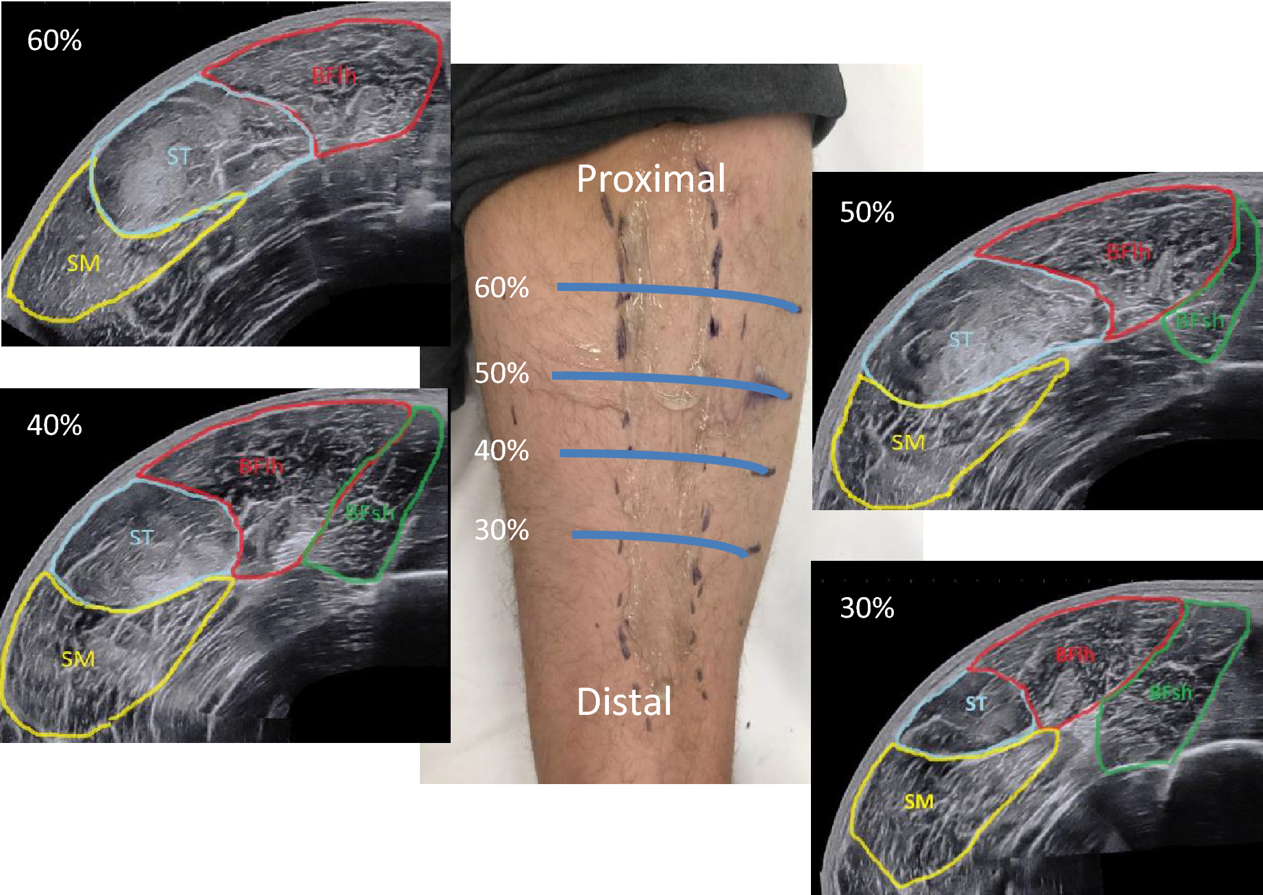
Panoramic ultrasound vs. MRI for the assessment of hamstrings cross-sectional area and volume in a large athletic cohort
Recomendado para você
-
Structural Characterization and Immunostimulatory Activity of a Homogeneous Polysaccharide from Sinonovacula constricta22 janeiro 2025
-
![Scorpion Battery Lock Strap Set (3) (Small) [SCP-LKSTRAPS] - HobbyTown](https://images.amain.com/cdn-cgi/image/f=auto,width=950/images/large/scp/scp-lkstraps_1.jpg) Scorpion Battery Lock Strap Set (3) (Small) [SCP-LKSTRAPS] - HobbyTown22 janeiro 2025
Scorpion Battery Lock Strap Set (3) (Small) [SCP-LKSTRAPS] - HobbyTown22 janeiro 2025 -
 SCP-007-J, Wiki Fundação SCP22 janeiro 2025
 -
 SCP's I made Minecraft Collection22 janeiro 2025
 -
 David Altmejd, Pyramid (2019)22 janeiro 2025
 -
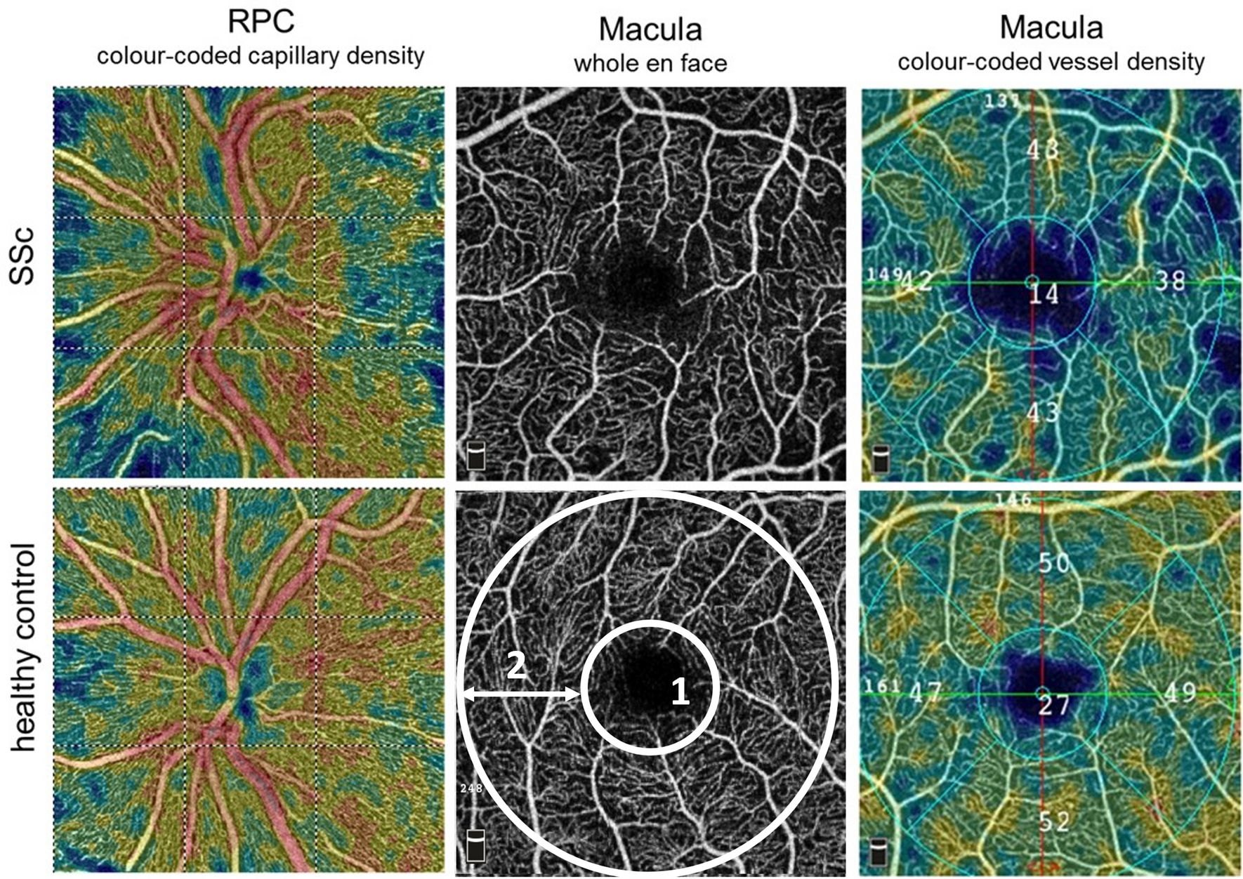 Altered ocular microvasculature in patients with systemic sclerosis and very early disease of systemic sclerosis using optical coherence tomography angiography22 janeiro 2025
 -
SCP Foundation22 janeiro 2025
-
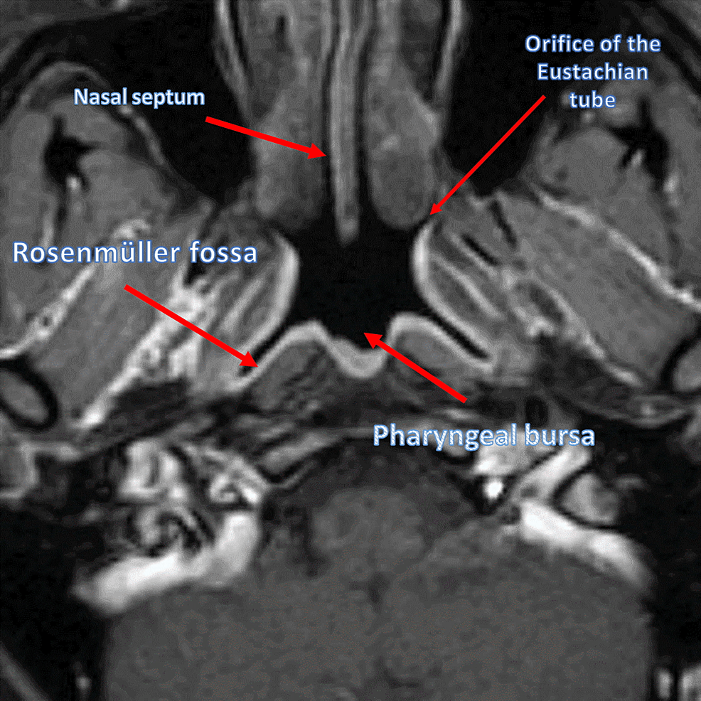 Cureus, Obstructive Sleep Apnea and Role of the Diaphragm22 janeiro 2025
 -
 Pathogenesis of neural tube defects: The regulation and disruption of cellular processes underlying neural tube closure - Engelhardt - 2022 - WIREs Mechanisms of Disease - Wiley Online Library22 janeiro 2025
 -
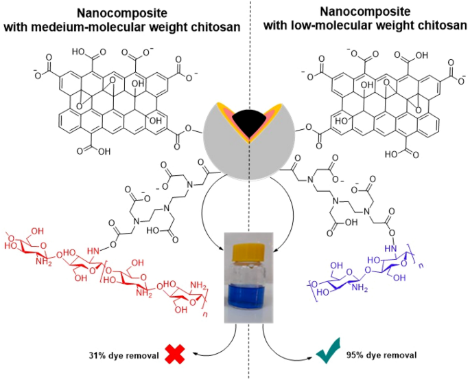 Magnetic Nanocomposites Containing Low and Medium-Molecular Weight Chitosan for Dye Adsorption: Hydrophilic Property Versus Functional Groups22 janeiro 2025
você pode gostar
-
Steven universo episodios novos22 janeiro 2025
-
/cdn.vox-cdn.com/uploads/chorus_image/image/62884927/RE2_Dec_Screen_10.0.jpg) Resident Evil 2 review: The new world of survival horror - Polygon22 janeiro 2025
Resident Evil 2 review: The new world of survival horror - Polygon22 janeiro 2025 -
 Gordon Hayward after injury: 'Is this my career?22 janeiro 2025
 -
 Rick Roll QR Code Wifi Sign Prank : Office Products22 janeiro 2025
 -
 Como consultar uma Inscrição Estadual ou CNPJ no Cadastro Centralizado de Contribuinte (CCC)?22 janeiro 2025
 -
Neil Druckmann on Instagram: Holy moly… This thing is so rad22 janeiro 2025
-
 Nintendo Switch Skyline Android Emulator Gets Killed Off With More Nintendo Switch Emulators To Follow? - Gameranx22 janeiro 2025
 -
Do any of the Straw Hat crew members have a bounty? - Quora22 janeiro 2025
-
 Pokémon X Rider FV 'Pikachu' - Puma - 387688 01 - empire yellow22 janeiro 2025
 -
 Scared Smiley Symbols & Emoticons22 janeiro 2025


